Raman Spectroscopy
Description
Light Scattering
Light falling on an object can be transmitted, reflected, absorbed, or scattered. In normal circumstances, only a very small part of the incident light is scattered by the object, and Raman spectroscopy utilizes only a small fraction of this scattered light.
There are two different possibilities for light scattering. The biggest part of the scattered light does not change its wavelength in the process and such scattering is known as elastic scattering or Rayleigh scattering as it was discovered and investigated by Lord Rayleigh. A small portion of the scattered light does change its wavelength in the process. This scattering was discovered by Sir C. V. Raman and it is thus known as Raman scattering or inelastic scattering.

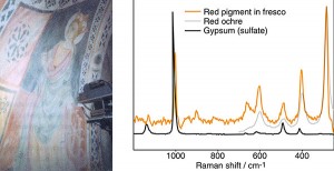
Example of Raman Spectra
Raman Scattering
The wavelength of the scattered light changes because the energy of the incident light causes changes in the vibration of the molecule or ion in the sample. The frequencies of such vibrations are dependent on the structure and thus each molecule or ion scatters the incident light in a different way. This fact can be used for the identification of the investigated samples such as pigments in a painting.
The intensity of the scattered light is plotted against its frequency and the result is a Raman spectrum of the sample. The spectra of the pigments had been measured and can be found in databases on the Internet (1)
The intensity of the scattered light is plotted against its frequency and the result is a Raman spectrum of the sample. The spectra of the pigments had been measured and can be found in databases on the Internet (1).
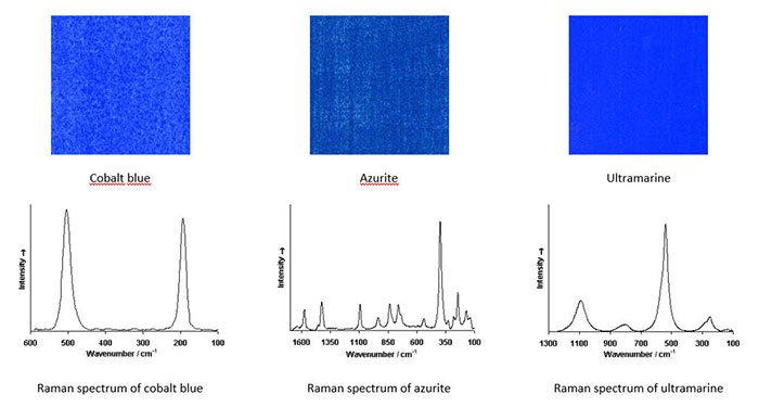
Examples of Raman Spectra of Pigments
The image below shows Raman spectra of three different blue pigments and it can be seen that all three spectra are distinctly different so that unequivocal identification of these pigments in a painting could easily be achieved.

Modern Developments
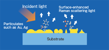
One solution for this disadvantage can be achieved by depositing the sample on a nanostructured metallic surface such as gold, copper, or silver. The intensity of Raman scattering, in this case, increases 104 to 108 times (ten thousand to hundred million times). This modern variant of Raman spectroscopy is known as Surface-Enhanced Raman Spectroscopy (SERS).

References
(1) Howell G. M. Edwards, John M. Chalmers, Raman Spectroscopy in Archaeology and Art History, Royal Society of Chemistry, 2005,
(2) Centeno S.A, Identification of artistic materials in paintings and drawings by Raman spectroscopy: Some challenges and future outlook, Journal of Raman Spectroscopy-1-47 (2016).
Procedure
Video: 'How to Do a Raman Spectrum' by HORIBA Scientific
Databases of Raman Spectra
Ian M. Bell, Robin J.H. Clark and Peter J. Gibbs, Raman Spectroscopic Library of Natural and Synthetic Pigments, Christopher Ingold Laboratories, University College London.
Ian M. Bell, Robin J.H. Clark, Peter J. Gibbs, Raman spectroscopic library of natural and synthetic pigments (pre- ≈ 1850 AD), Spectrochimica Acta Part A: Molecular and Biomolecular Spectroscopy, Volume 53, Issue 12, 15 October 1997, Pages 2159–2179.
Burgio L. and Clark R. Library of FT-Raman spectra of pigments, minerals, pigment media and varnishes, and supplement to existing library of Raman spectra of pigments with visible excitation, Spectrochimica Acta – Part A: Molecular and Biomolecular Spectroscopy (2001) 57(7) 1491-1521
DOI: 10.1016/S1386-1425(00)00495-9
Pozzi, F., Lombardi, J. R., & Leona, M. (2013). Winsor & Newton original handbooks: a surface-enhanced Raman scattering (SERS) and Raman spectral database of dyes from modern watercolor pigments. Heritage Science, 1(1), 23. doi:10.1186/2050-7445-1-23
P. Vandenabeele1, L. Moens, H. G. M. Edwards and R. Dams, Raman spectroscopic database of azo pigments and application to modern art studies, Journal of Raman Spectroscopy, Volume 31, Issue 6, pages 509–517, June 2000.
Nadim C. Scherrer a, Zumbuehl Stefan, Delavy Francoise, Fritsch Annette, Kuehnen Renate, Synthetic organic pigments of the 20th and 21st century relevant to artist’s paints: Raman spectra reference collection, Spectrochimica Acta Part A 73 (2009) 505–524. Available as pdf.
IRUG (Infrared and Raman User Group) Spectral Database
Marucci, G., A. Beeby, A. W. Parker, and C. E. Nicholson. “Raman Spectroscopic Library of Medieval Pigments Collected with Five Different Wavelengths for Investigation of Illuminated Manuscripts.” Analytical Methods 10, no. 10 (n.d.): 1219–36. doi:10.1039/C8AY00016F.
Examples of Use
Diego Velázquez
P.C. Gutiérrez-Neiraa, F. Agulló-Ruedab, A. Climent-Fonta, C. G. Raman spectroscopy analysis of pigments on Diego Velázquez paintings. Vibrational Spectroscopy, 69, 2013, 13– 20.
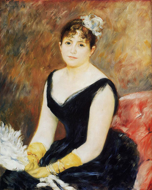
Pierre-Auguste Renoir, Portrait of Mme Léon Clapisson, 1883
The scientists found a vivid scarlet coloured pigment in the areas of the painting hidden behind the frame where it was protected from light. They then identified this pigment as carmine by Surface Enhanced Raman Spectroscopy (SERS). (The colours in the image below are not entirely correct. For the image in correct colours see the website of the Art Institute of Chicago.

The data from this analysis have been employed to construct a digitally rejuvenated version of the painting and we can now see how the painting could have looked at the time of its creation. For more information on the method of digital rejuvenation of paintings in general and for the details on the digital rejuvenation of this particular painting see the page Digital Restoration of Paintings on this website.
Renoir’s True Colors: Science Solves a Mystery
19th and 20th Century Works of Art
Identification of Specific Pigments
Hannah E. Mayhew, David M. Fabian, Shelley A. Svobodab and Kristin L. Wustholz, Surface-enhanced Raman spectroscopy studies of yellow organic dyestuffs and lake pigments in oil paint, Analyst, 2013,138, 4493-4499. DOI: 10.1039/C3AN00611E.
Robin J. H. Clark, Lucas Cridland, Benson M. Kariuki, Kenneth D. M. Harris and Robert Withnall, Synthesis, structural characterisation and Raman spectroscopy of the inorganic pigments lead tin yellow types I and II and lead antimonate yellow: their identification on medieval paintings and manuscripts. J. Chem. Soc., Dalton Trans., 1995, 2577-2582. DOI: 10.1039/DT9950002577.
A. Nevin, et al., Advances in the analysis of red lake pigments from 15th and16th c. paintings using fluorescence and Raman spectroscopy, Int. Conf. on non-destructive investigations and microanalysis for the diagnostics and conservation of cultural and environmental heritage (ART 2011), 13-15 April 2011, Florence, Italy.
Pozzi, F., van den Berg, K. J., Fiedler, I. and Casadio, F. (2014), A systematic analysis of red lake pigments in French Impressionist and Post-Impressionist paintings by surface-enhanced Raman spectroscopy (SERS). J. Raman Spectrosc.. doi: 10.1002/jrs.4483.
Whitney, A. V., et al. An innovative surface-enhanced Raman spectroscopy (SERS) method for the identification of six historical red lakes and dyestuffs. Journal of Raman Spectroscopy 37(10), 2006, 993–1002. Available as pdf.
Osticioli, I., Mendes, N. F. C., Nevin, A., Gil, F. P. S. C., Becucci, M., & Castellucci, E. Analysis of natural and artificial ultramarine blue pigments using laser induced breakdown and pulsed Raman spectroscopy, statistical analysis and light microscopy. Spectrochimica Acta. Part A, Molecular and Biomolecular Spectroscopy, 73(3), (2009) 525–31. doi:10.1016/j.saa.2008.11.028.
R.J.H. Clark, M.L. Franks, The resonance Raman spectrum of ultramarine blue, Chemical Physics Letters
Volume 34, Issue 1, 1 July 1975, Pages 69-72.
Alessia Coccato, Jan Jehlicka, Luc Moens and Peter Vandenabeele, Raman spectroscopy for the investigation of carbon-based black pigments, Journal of Raman Spectroscopy, Special Issue: 11th International GeoRaman Conference, Volume 46, Issue 10, pages 1003–1015, October 2015. DOI: 10.1002/jrs.4715. Available as pdf.
Eugenia P. Tomasini, Emilia B. Halac, María Reinoso, Emiliano J. Di Liscia and Marta S. Maier, Micro-Raman spectroscopy of carbon-based black pigments, Journal of Raman Spectroscopy, Special Issue: Raman spectroscopy in art and archaeology, Volume 43, Issue 11, pp. 1671–1675, November 2012.
F. Casadio1, A. Bezúr, I. Fiedler1, K. Muir T. Trad and S. Maccagnola, Pablo Picasso to Jasper Johns: a Raman study of cobalt-based synthetic inorganic pigments, Special Issue: Raman spectroscopy in art and archaeology, Volume 43, Issue 11, pages 1761–1771, November 2012.
Video: 'Science meets art: investigating pigments in art and archaeology' by R. Clark
Further Reading
References
(1) Robin J. H. Clark, Raman Microscopy in the Identification of Pigments on Manuscripts and Other Artwork. In Scientific Examination of Art: Modern Techniques in Conservation and Analysis, Arthur M. Sackler Colloquium on Scientific Examination of Art, National Academy of Sciences (U.S.), National Academies Press, 2005, 162-185. (pdf).
(2) Centeno S.A, Identification of artistic materials in paintings and drawings by Raman spectroscopy: Some challenges and future outlook, Journal of Raman Spectroscopy-1-47 (2016).
(3) Natalia Navas, Julia Romero-Pastor, Eloisa Manzanoaand, Carolina Cardell, Raman spectroscopic discrimination of pigments and tempera paint model samples by principal component analysis on first-derivative spectra, Journal of Raman Spectroscopy (Impact Factor: 2.67). 11/2010; 41(11):1486 – 1493. DOI: 10.1002/jrs.2646.
(4) M. Castanys, M. J. Soneira, and R. Perez-Pueyo, Automatic Identification of Artistic Pigments by Raman Spectroscopy Using Fuzzy Logic and Principal Component Analysis, Laser Chemistry, Volume 2006, Article ID 18792, 8 pages.
(5) Howell G. M. Edwards, John M. Chalmers, Raman Spectroscopy in Archaeology and Art History, Royal Society of Chemistry, 2005.
(6) M.C. Chillón, Perla Ferrer, Sergio Ruiz-Moreno, Alejandro López-Gil, Avoiding frauds: Raman spectroscopy, a fundamental tool in the analysis of artworks. Proceedings of the 3th International Conference on Heritage and Sustainable Development HERITAGE 2012, Oporto
