IR Spectroscopy
IRDescription
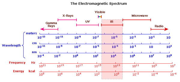
Infrared spectroscopy is an analytical method for the identification of pigments in paintings. The pigment sample is irradiated by infrared radiation of wavelength between around 1000 and 4000 nm (nanometers, 1 nm = 1 billionth of a meter) as can be seen in the image below.
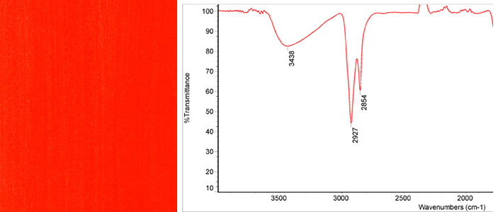
Infrared Spectra
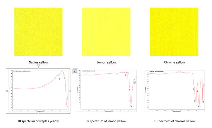
Parts of the radiation is absorbed by the sample due to the vibrations of the molecules or ions. Such vibrations are dependent on the structure of the molecule and thus are unique to the particular chemical substance. The intensity of the reflected or transmitted IR-radiation is plotted against its wavelength and such a plot is called the infrared spectrum. IR-spectra can be found in several databases on the Internet for the great number of pigments. The image below shows three optically similar yellow pigments with distinctly different IR-spectra.
Modern Developments
Later developments made it possible to record the same spectrum many times over in a very short time and thus greatly improve the quality of the spectra. The mathematical transformation needed to record the spectrum several times over is called Fourier transformation (FT-IR-Spectroscopy).
The latest generation of instruments allows recording reflection spectra of paint samples. With such instruments, it is not necessary to extract a paint sample from the painting and place it in the sample holder of the spectrometer. The spectra can be recorded contactless by placing the instrument in front of the painting. This makes the method non-destructive and thus greatly enhances its potential (1).
References
(1) Bruker Optical Inc., Contactless analysis of paintings with FT-IR spectroscopy, Application Note AN #95.
Electromagnetic Spectrum
Databases of IR-Spectra
Database of ATR-FT-IR Spectra of Pigments, Institute of Chemistry, University of Tartu, Estonia
Vahur, S., University of Tartu, Estonia, Database of ATR-IR spectra of materials related to paints and coatings. This database contains IR-spectra of pigments mixed with linseed oil.
Carlos Eduardo Silva, Luciana P Silva, H. G. M. Edwards, Luiz Fernando Cappa De Oliveira, Diffuse reflection FTIR spectral database of dyes and pigments, Anal Bioanal Chem (2006) 386:2183–2191DOI 10.1007/s00216-006-0865-8. Available as pdf.
Procedure
Video: How to Use an IR Spectrometer and Sample Prep' by Supreme Science
Video: 'IR-Spectroscopy' by Royal Society of Chemistry
Examples of Use
Henri Matisse, The Joy of Life, 1905-06
E. Pouyet , M. Cotte, B. Fayard, M. Salomé, F. Meirer, A. Mehta, E. S. Uffelman, A. Hull, F. Vanmeert and 5 more, 2D X-ray and FTIR micro-analysis of the degradation of cadmium yellow pigment in paintings of Henri Matisse, Applied Physics A, November 2015, Volume 121, Issue 3, pp 967-980.
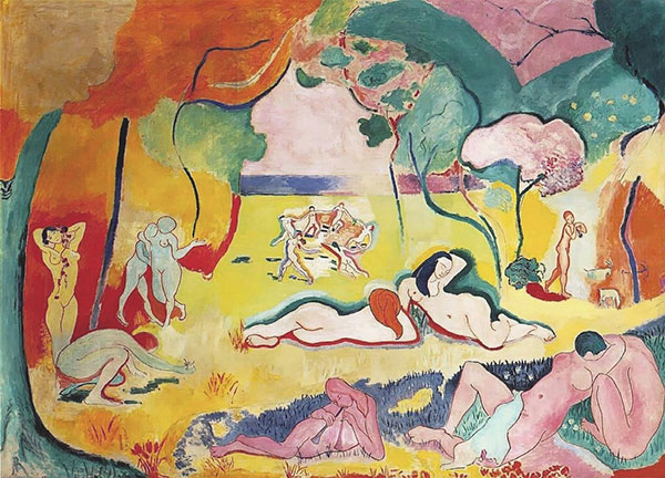
(Image: Licensed under PD-US via Wikipedia, Image URL)
Abstract of the above publication
“The chemical and physical alterations of cadmium yellow (CdS) paints in Henri Matisse’s The Joy of Life (1905–1906, The Barnes Foundation) have been recognized since 2006, when a survey by portable X-ray fluorescence identified this pigment in all altered regions of the monumental painting. This alteration is visible as fading, discoloration, chalking, flaking, and spalling of several regions of light to medium yellow paint. Since that time, synchrotron radiation-based techniques including elemental and spectroscopic imaging, as well as X-ray scattering have been employed to locate and identify the alteration products observed in this and related works by Henri Matisse. This information is necessary to formulate one or multiple mechanisms for degradation of Matisse’s paints from this period, and thus ensure proper environmental conditions for the storage and the display of his works. This paper focuses on 2D full-field X-ray Near Edge Structure imaging, 2D micro-X-ray Diffraction, X-ray Fluorescence, and Fourier Transform Infra-red imaging of the altered paint layers to address one of the long-standing questions about cadmium yellow alteration—the roles of cadmium carbonates and cadmium sulphates found in the altered paint layers. These compounds have often been assumed to be photo-oxidation products, but could also be residual starting reagents from an indirect wet process synthesis of CdS. The data presented here allow identifying and mapping the location of cadmium carbonates, cadmium chlorides, cadmium oxalates, cadmium sulphates, and cadmium sulphides in thin sections of altered cadmium yellow paints from The Joy of Life and Matisse’s Flower Piece (1906, The Barnes Foundation). Distribution of various cadmium compounds confirms that cadmium carbonates and sulphates are photo-degradation products in The Joy of Life, whereas in Flower Piece, cadmium carbonates appear to have been a [(partially) unreacted] starting reagent for the yellow paint, a role previously suggested in other altered yellow paints.”
Vincent van Gogh, La Mousmé, 1888
John K. Delaney, Elizabeth Walmsley, Barbara H. Berrie, and Colin F. Fletcher, Multispectral Imaging of Paintings in the Infrared to Detect and Map Blue Pigments, In Sackler NAS Colloquium, Scientific Examination of Art: Modern Techniques in Conservation and Analysis (2005), pp. 120-136. Available as pdf.
Abstract from the above publication
“In this paper spectral imaging in the reflective infrared (IR) spectral region (0.7 to 2.5 microns) is examined for its potential to discriminate and identify blue pigments in paintings. The blue pigments considered are azurite, indigo, Prussian blue, cobalt blue, ultramarine, and phtalocyanine blue. The blue areas in this painting by Van Gogh have been investigated by the authors and the pigments identified as prussian blue and artificial ultramarine.”
Identification of Individual Pigments
Database of ATR-FT-IR Spectra of Pigments, Institute of Chemistry, University of Tartu, Estonia
Vahur, S., University of Tartu, Estonia, Database of ATR-IR spectra of materials related to paints and coatings. This database contains IR-spectra of pigments mixed with linseed oil.
Signe Vahur, Anu Teearu, Ivo Leito, ATR-FT-IR spectroscopy in the region of 550–230 cm−1 for identification of inorganic pigments, Spectrochimica Acta Part A 75 (2010) 1061–1072.
Carlos Eduardo Silva, Luciana P Silva, H. G. M. Edwards, Luiz Fernando Cappa De Oliveira, Diffuse reflection FTIR spectral database of dyes and pigments, Anal Bioanal Chem (2006) 386:2183–2191DOI 10.1007/s00216-006-0865-8. Available as pdf.
NICODOM FTIR Library Dyes and Pigments (1400 spectra)
Buti D, Rosi F, Brunetti BG, Miliani C., In-situ identification of copper-based green pigments on paintings and manuscripts by reflection FTIR. Anal Bioanal Chem. 2013 Mar;405(8):2699-711. doi: 10.1007/s00216-013-6707-6. Epub 2013 Jan 23.
Eglė Gražėnaitė, Jonas Kiuberis, Aivaras Kareiva, Aldona Beganskienė, Jūratė Senvaitienė, XRD and FTIR characterisation of historical green pigments and their lead-based glazes, chemija. 2014. vol. 25. No. 4. P. 199–205.
Helwig, K. (2017). 11. The characterisation of iron earth pigments using infrared spectroscopy. irug.org, [online] p. Available at: https://www.academia.edu/1006437/11._The_characterisation_of_iron_earth_pigments_using_infrared_spectroscopy?auto=download [Accessed 25 Jul. 2017].
Further Reading
References
(1) Derrick, M.R., Stulik, D. and Landry J.M., Infrared Spectroscopy in Conservation Science, Scientific Tools for Conservation, Getty Publications, 2000. Available in pdf format.
(2) Richard Newman, Some applications of infrared spectroscopy in the examination of painting materials, Journal of the American Institute for Conservation (JAIC) 1979, Volume 19, Number 1, Article 6 (pp. 42 to 62). Available online.
(3) John K. Delaney, Mathieu Thoury, Jason G. Zeibel et al., Visible and infrared imaging spectroscopy of paintings and improved reflectography, Heritage Science (2016), 4:6, https://doi.org/10.1186/s40494-016-0075-4
(4) Francesca Casadio, Lucia Toniolo, The analysis of polychrome works of art: 40 years of infrared spectroscopic investigations, Journal of Cultural Heritage, Volume 2, Issue 1, 2001, Pages 71-78. https://doi.org/10.1016/S1296-2074(01)01107-4