X-Ray Radiography
XRRDescription of X-Ray Radiography
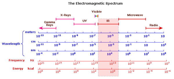
X-ray radiography is one of the oldest and most widely employed scientific methods for the investigation of works of art. X-rays are electromagnetic waves with a very short wavelength in the range of 1 to 250 pm, where 1 pm = 10-12 m (see the image of the electromagnetic spectrum below). They are able to penetrate many materials and thus allow an insight into the inner layers of a painting.
The transmission of X-rays through any material is inversely proportional to the atomic weight of the elements in the sample. This means that only relatively heavy elements such as mercury or lead can stop the x-rays sufficiently to be visible in a radiograph. Thus only pigments containing these elements can contribute to a radiograph image. The choice of pigments containing these elements is correspondingly limited: lead white, lead-tin-yellow, and vermilion (mercuric sulfide) being the most frequently used ones.

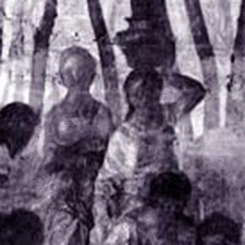
G. Bellini, Feast of Gods, Detail
X-Ray Image
Electromagnetic Spectrum
Procedure for X-Ray Radiography
The painting is placed on a sheet of photographic film and irradiated by X-rays. The layers of the painting absorb a portion of the X-rays and the remaining x-rays form an image on the film. A modern variant of the method is shown in the following video by ntb X-ray imaging, where an X-ray detector is used instead of a photographic film.
Modern Developments
One of the more recent developments in the area of X-ray radiography is a computational method for removing the disturbing traces of the wooden support (cradle) of the frame from an x-ray radiograph. Platypus is a software solution that comes both as a standalone application and a Photoshop plugin. It is specifically designed to digitally remove cradling artifacts in X-ray images of paintings on a panel (2).
Video: 'X-Ray Radiography' by ntb X-Ray Imaging
References
(1) M. Schreiner, B. Frühmann, D. Jembrih-Simbürger, R. Linke, X-rays in Art and Archeology – an Overview, International Centre for Diffraction Data 2004, Advances in X-ray Analysis, Volume 47, presented at the Denver X-ray Conference (DXC) on Applications of X-ray Analysis.
(2) R. Yin, D. Dunson, B. Cornelis, B. Brown, N. Ocon and I. Daubechies, Digital Cradle Removal in X-ray Images of Art Paintings, IEEE International Conference on Image Processing 2014 (ICIP2014). Available as pdf.
Examples of Use
Jan van Eyck, The Ghent Altarpiece, 1430-32
Website of the project “Closer to Van Eyck, Rediscovering the Ghent Altarpiece“. The entire altarpiece was investigated by IR-Reflectography and X-Ray Radiography. There are also zoomable high-resolution images in visible light and other information on this artwork. Both images are courtesy of this website.
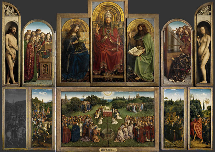
Jan van Eyck, The Ghent Altarpiece, 1430-32, visible light

Raphael, Portrait of Pope Julius II, 1511
The painting in its present state at the National Gallery London.
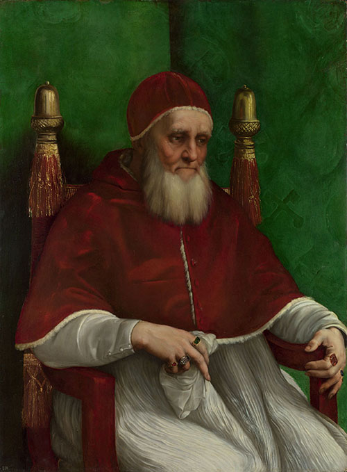
The X-ray investigation of this painting at the National Gallery London has revealed, amongst other new insights, that the green background has been added later by Raphael. The original background shows a series of heraldic emblems.
The very thorough examinations of this masterwork by several scientific methods such as IR reflectography and microscopy are extensively documented on the website of the Raphael Research Resource.
References
(1) Dunkerton, Jill, and Ashok Roy. “The altered background of Raphael’s portrait of Pope Julius II in the National Gallery, London”. The Burlington Magazine 146 (2004) 757-759.
(2) A. Roy, ‘Portrait of Pope Julius II’, The Mellon Digital Documentation Project, 2008.
G. Bellini and Titian, The Feast of the Gods, 1514-29
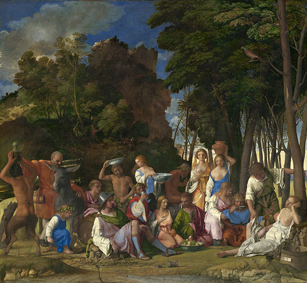
New Insights From the X-Ray Investigation
The extensive investigation at the National Gallery of Art in Washington is thoroughly described on the page for this painting on this website. X-ray radiographs revealed several striking insights into the genesis of the painting.
It came to light, that Bellini changed the composition in the foreground slightly, one example being the eroticization of the female figures. The blue dress of the nymph with the jar on her head was originally placed high on her chest. Bellini then overpainted it so that her right breast is exposed in the present version. The original less erotic version can clearly be seen in the detail of the X-radiograph.
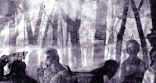
The X-ray radiograph of the middle portion of the painting shows that the background behind the figures in the foreground originally consisted of single tree trunks, similar to those still visible on the right side of the painting. It is now known that the left part of the background was overpainted after Bellini’s death by Titian. For a more detailed account see the page on this painting on this website and the references below.
References
(1) Plesters, J. The Feast of the Gods: Conservation, Examination, and Interpretation. Studies in the History of Art, 40, 1990.
(2) Plesters, J., Examination of Giovanni Bellini’s Feast of the Gods: A Summary and Interpretation of the Results, Studies in the History of Art, Vol. 45, Symposium Papers XXV: Titian 500 (1993), pp. 374-391
(3) Bull, D., The Feast of the Gods: Conservation and Investigation, Studies in the History of Art, Vol. 45, Symposium Papers XXV: Titian 500 (1993), pp. 366-373
(4) Investigating Bellini’s Feast of the Gods, WebExhibits website.
(5) John Walker, Bellini and Titian at Ferrara: A Study of Styles and Taste, Phaidon Publishers, 1956
Titian, The Death of Actaeon, 1555-75
The X-ray investigation of this painting at the National Gallery London has revealed several changes made by Titian in the course of his work.
Further Reading
References
(1) Murray Pease, Note on the Radiography of Paintings, Bulletin of the Metropolitan Museum of Art, available as pdf.
(2) Janssens, K., Van der Snickt, G., Vanmeert, F. et al. Non-Invasive and Non-Destructive Examination of Artistic Pigments, Paints, and Paintings by Means of X-Ray Methods. Top Curr Chem (Z) 374, 81 (2016) doi:10.1007/s41061-016-0079-2
(3) Gilardoni, Arturo, Orsini Riccardo. Ascani, and Silvia Taccani. X-rays in Art. Mandello Lario: Gilardoni, 1977.
(4) Lang, Janet, and Andrew Middleton. Radiography of Cultural Material. 2nd ed. Burlington: Butterworth-Heinemann, 2005.
(5) Graham, Daniel, and Thomas Eddie. X-ray Techniques in Art Galleries and Museums. Bristol: Adam Hilger Ltd, 1985.
(6) A. Cabal, O. Schalm, P. Eyskens, P. Willems, A. Harth and P. Van Espen, Comparison of x-ray absorption and emission techniques for the investigation of paintings, X-Ray Spectrometry, Volume 44, Issue 3, pages 141–148, May/June 2015. Presented at the European Conference on X-Ray Spectrometry, Bologna, Italy, 15–20 June 2014. DOI: 10.1002/xrs.2591.
(7) Richard Newman, Applications of x rays in art authentication: radiography, x-ray diffraction, and x-ray fluorescence, Proc. SPIE 3315, Scientific Detection of Fakery in Art, 31 (May 25, 1998); doi:10.1117/12.308590; http://dx.doi.org/10.1117/12.308590. From: Walter McCrone; Duane R. Chartier; Richard J. Weiss, Conference Volume 3315 Scientific Detection of Fakery in Art, San Jose, CA | January 24, 1998.
(8) Padfield, J., Saunders, D., Cupitt, J., Atkinson, R. ‘Improvements in the Acquisition and Processing of X-ray Images of Paintings’. National Gallery Technical Bulletin Vol 23, pp 62–75. Available as pdf.
(9) Kress Technical Art History Website, X-Ray Radiography, [online] Available at https://www.artcons.udel.edu/outreach/kress/examination-methods-and-scientific-terms/x-radiography [Accessed 9 Jul. 2017].
(10) (2) M. Schreiner, B. Frühmann, D. Jembrih-Simbürger, R. Linke, X-Rays in Art and Archeology – an Overview, International Centre for Diffraction Data 2004, Advances in X-ray Analysis, Volume 47, presented at the Denver X-ray Conference (DXC) on Applications of X-ray Analysis.