X-Ray Fluorescence
XRFDescription of X-Ray Fluorescence
In this method, the sample is irradiated by an intense and focused x-ray beam. The energy of the x-rays is sufficient to expel electrons from the inner shells (close to the atomic nucleus) in an atom. Electrons from outer shells fall subsequently into the holes left by the expelled electrons and emit x-rays in the process. As every atom has its own particular structure of its electron shells, the energy of the emitted x-rays is characteristic for each element and can be used for its identification.
X-Ray Fluorescence Spectra
The intensity of the emitted x-rays can be plotted against their energy. Such a plot is called an x-ray fluorescence spectrum. The image at the right shows such a spectrum for a multitude of elements. The signal for each element is registered at characteristic energy for that element.
The irradiation source can be a conventional x-ray generator or in the more recent applications, a synchrotron (particle accelerator) can also be employed as the source of a very intense x-ray beam. Radioactive materials emanating γ-rays can also be used for this method with the big advantage of being able to miniaturize the instruments to such an extent that they can be made handheld (portable XRF, p-XRF) (2).

Rembrandt, An Old Man in a Military Costume,1630-31
The painting being scanned in an XRF apparatus
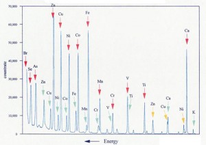
X-Ray Fluorescence spectra of various elements
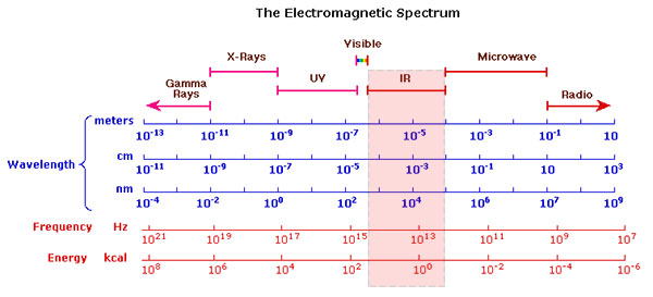
Electromagnetic Spectrum
Video: 'X-Ray Fluorescence Spectroscopy (XRF) Explained' by Captain Corrosion
Procedure
Pigment Identification
In conducting a measurement, a specific area of the painting is irradiated by x-rays and the intensity of the emitted x-rays is recorded. The resulting signals can be assigned to the specific elements and in turn, the elements can then be tentatively assigned to specific pigments in the context of the painting, e.g. sodium can be assigned to ultramarine, mercury to vermilion and copper to either malachite, azurite or verdigris. Pigment analysis of a small area of the painting can thus be obtained in this way. Bigger instruments make it possible to scan the whole painting in order to arrive at a complete pigment analysis for the entire painting.
Database of XRF-Spectra of Pigments, CHSOS website
Single-Element Distribution Maps
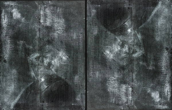
The XRF method offers also another even more interesting insight into the inner secrets of painting. Having the complete scan of the painting (as described above) one can then plot the intensity of the x-rays for each specific energy (and thus for each element) over the area of the entire painting. The resulting single-element maps show the distribution of a single pigment in the painting. The following images are examples of a recent x-ray fluorescence investigation of Rembrandt’s painting “An Old man in Military Costume“, 1630-31 (1). The face of a young man can clearly be distinguished in these XRF distribution maps for lead white and vermilion The underlying painting of the young man is rotated 180 degrees. The detailed description of this investigation is shown below under “Examples of use”.
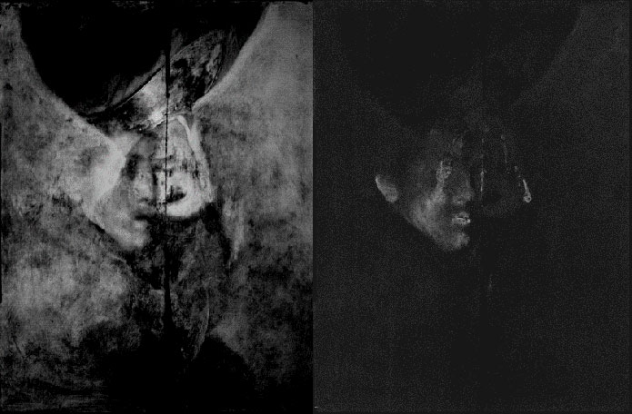
Rembrandt, An Old Man in Military Costume, 1630-31.
Single-element distribution maps for lead corresponding to the pigment lead white (left) and mercury corresponding to the pigment vermilion (right)
Images courtesy of The J. Paul Getty Trust (1).
Taking the above into account, one can see that XRF can be classified as a spectroscopic method and as an imaging method at the same time.
Video: 'The Non-Invasive Analysis of Painted Surfaces' by Nicolas Barbi
References
(1) Karen Trentelman, Koen Janssens, Geert van der Snickt, Yvonne Szafran, Anne T. Woollett, Joris Dik, Rembrandt’s An Old Man in Military Costume: the underlying image re-examined, Applied Physics A, November 2015, Volume 121, Issue 3, pp 801-811. Available as pdf.
(2) Bruker Corporation, How Handheld XRF Works: A Step-by-Step Guide.
Examples of Use
Rembrandt van Rijn, An Old Man in Military Costume, 1630-31
The Painting in Its Present State
The painting at The J. Paul Getty Museum
Video narrative about the painting from The Getty Museum.
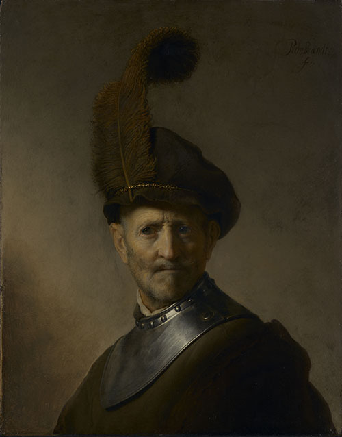
Rembrandt van Rijn, An Old Man in Military Costume, 1630-31
Conventional X-Ray Radiography
The painting was first investigated by conventional X-ray radiography and an underlying portrait of a young man was discovered beneath the visible image. The quality of the x-ray image was not sufficient to discern any details of the underlying portrait. It should be noted that Rembrandt rotated the canvas 180 degrees before he overpainted the old image with the ‘An Old Man in Military Costume‘.
X-radiographs of An Old Man in Military Costume. The X-ray at right is inverted to better show the hidden painting.
Image courtesy of The J. Paul Getty Trust.
Neutron Activation Radiography (NAAR) and X-Ray Fluorescence (XRF) Investigations (1,2)
The latest investigation by Trentelman et al. (2) employing both the x-ray fluorescence (XRF) and neutron activation autoradiography (NAAR) brought a considerable improvement in image quality and in the identification of pigments as well. The NAAR images showed improved detail in the face and the cloak of the young man and the XRF data made it possible to obtain relatively clear images of the underlying painting and roughly identify the pigments used by Rembrandt in the underlying painting as well (1). The images below are single-element maps for the two elements lead and mercury. Lead corresponds to the pigment lead white and mercury is contained in the red pigment vermilion.
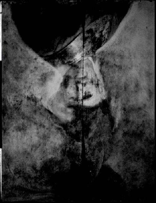
Rembrandt, Old Man in Military Costume, 1630-31. XRF single-element image map for lead rotated 180 degrees.
Image courtesy of J. Paul Getty Trust.
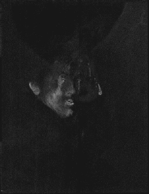
Rembrandt, Old Man in Military Costume, 1630-31. XRF single-element image map for mercury rotated 180 degrees.
Image courtesy of J. Paul Getty Trust.
Digital Reconstruction
The results of the entire investigation using both NAAR and XRF methods could then be converted into a colour digital reconstruction of the underlying portrait of the young man. Comparing this image to the conventional x-ray images above shows the impressive extent of the technical development of the scientific investigation methods in the last decades.
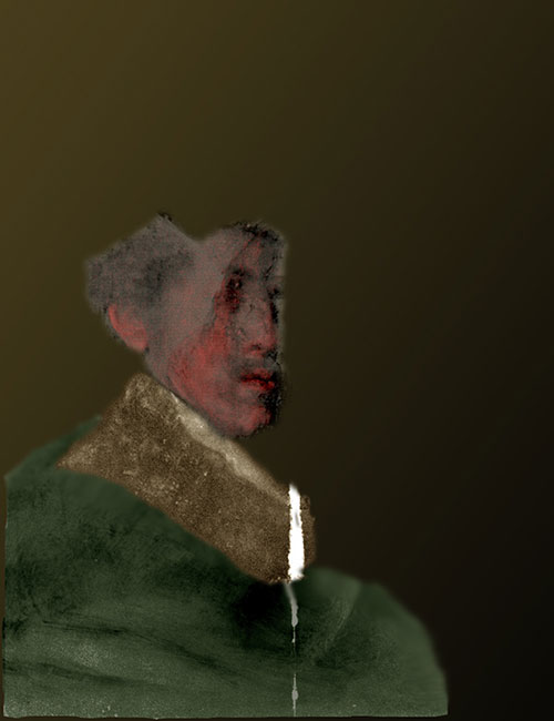
Digital reconstruction of the underlying portrait of the young man.
Image courtesy of The J. Paul Getty Trust.
References
(1) Annelisa Stephan, A Hidden Rembrandt Has Been Digitally Reconstructed in Color, IRIS – The online magazine of The Getty, 1 September 2015.
(2) Karen Trentelman, Koen Janssens, Geert van der Snickt, Yvonne Szafran, Anne T. Woollett, Joris Dik, Rembrandt’s An Old Man in Military Costume: the underlying image re-examined, Applied Physics A, November 2015, Volume 121, Issue 3, pp 801-811. Available as pdf.
Paolo Veronese
Erich Stuart Uffelman, Elizabeth Court, John Marciari, Alexis Miller, and Lauren Cox, Handheld XRF Analyses of Two Veronese Paintings, Collaborative Endeavors in the Chemical Analysis of Art and Cultural Heritage Materials, Chapter 3, pp 51–73. Available as pdf.
Abstract of the above paper
“Paolo Veronese’s painting of Apollo and Daphne at the San Diego Museum of Art was suspected of containing large areas of degraded smalt (a pigment derived from ground cobalt glass) in the sky, causing the original blue color to have turned gray. Handheld XRF analysis of the painting confirmed the presence of cobalt in all spots involving the sky. For purposes of contrast, Veronese’s painting of Madonna and Child with St. Elizabeth, the Infant St. John, and St. Catherine at The Timken Museum of Art was analyzed by handheld XRF. The Timken’s Veronese’s sky is still blue, and copper (almost certainly present in the form of azurite) was found instead of cobalt.”
Edgar Degas, Portrait of a Woman, 1876-80
A hidden portrait was found beneath the surface of the painting by the researchers at the National Gallery of Victoria, Melbourne, Australia, and the Australian Synchrotron Facility (1).
The existence of the “hidden” portrait was known for a long time because Degas painted the second portrait directly over the first one using thin paint of low opacity. This makes it possible to discern the traces of the first portrait by the naked eye.
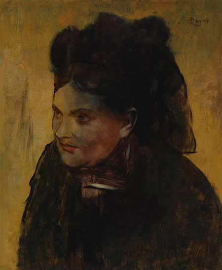
Edgar Degas, Portrait of a Woman, 1876-80
Image courtesy of reference (1)
A conventional x-ray image allows the first glimpse of the underlying portrait.
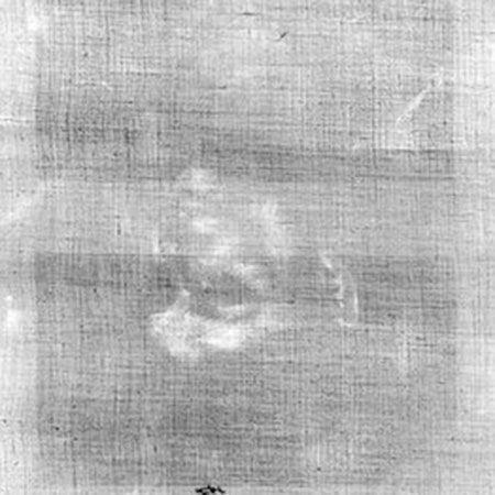
Conventional x-ray image
Image courtesy of reference (1)
The painting was then irradiated by high energy x-rays using a cyclotron as the x-ray source. The resulting x-ray fluorescence was recorded by scanning the whole painting at all wavelengths corresponding to elements found in the usual pigments. The result was elemental distribution maps for each element as shown in the image below.

The painting is prepared for the scanning XRF investigation
Image courtesy of reference (1)
The resulting elemental distribution maps are shown in the next image.
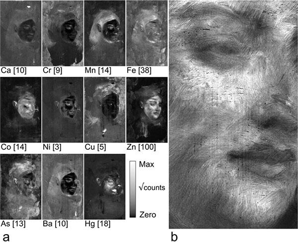
a) Elemental distribution maps, b) enlarged elemental map for zinc (Zn)
Image courtesy of reference (1)
The elemental maps can now be tentatively assigned to the corresponding pigments. The element zinc (Zn) is contained in zinc white employed in the highlights of the face, the red pigment vermilion employed for the lips and the left cheek contains mercury (Hg), and both manganese (Mn) and iron (Fe) are constituents of the brown pigment umber used for the hair.
In the last step, the information on the pigment distribution together with the assignment of the proper color to each pigment can now be used to construct a digital color image of the underlying painting.
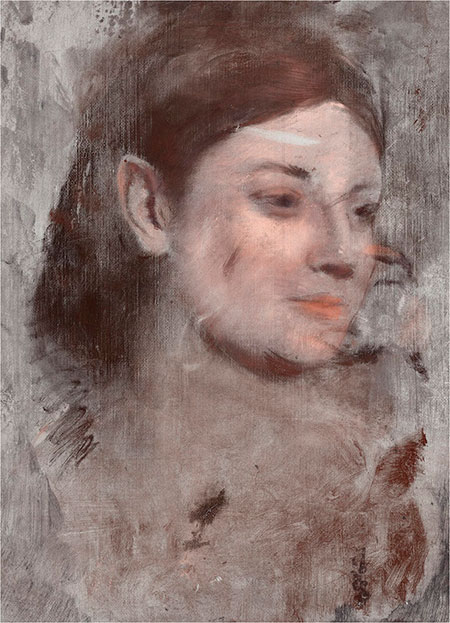
Digital reconstruction of the underlying portrait
Image courtesy of reference (1)
The quality and detail of this image are indeed stunning, especially if compared to the classical x-ray image shown above.
References
(1) David Thurrowgood, David Paterson, Martin D. de Jonge, Robin Kirkham, Saul Thurrowgood & Daryl L. Howard, A Hidden Portrait by Edgar Degas, Nature, Scientific Reports volume 6, Article number: 29594 (2016), doi:10.1038/srep29594
Vincent van Gogh, Grass, 1887
Painting at the Kröller-Müller Museum in Otterlo, The Netherlands
Joris Dik, Koen Janssens, Geert Van Der Snickt, Luuk van der Loeff, Karen Ricker and Marine Cotte, Visualization of a Lost Painting by Vincent van Gogh Using Synchrotron Radiation Based X-ray Fluorescence Elemental Mapping, Anal. Chem., 2008, 80 (16), pp 6436–6442, DOI: 10.1021/ac800965g.
Anila Anitha, Andrei Brasoveanu, Marco Duarte, Shannon Hughes, Ingrid Daubechies, Joris Dik, Koen Janssens, Matthias Alfeld, Restoration of X-ray fluorescence images of hidden paintings, Signal Processing 93(3), 592–604, March 2013, Available online.
Article at the website of DESY
Pigment Characterization and Detection
Koen Janssens, Matthias Alfeld, Geert Van der Snickt, Wout De Nolf, Frederik Vanmeert, Marie Radepont, Letizia Monico, Joris Dik, Marine Cotte, Gerald Falkenberg, Costanza Miliani, and Brunetto G. Brunetti, The Use of Synchrotron Radiation for the Characterization of Artists’ Pigments and Paintings, Annual Review of Analytical Chemistry, (2008) 6(1):399-425 · June 2013. Available as pdf.
Quote from the abstract of the above publication:
We review methods and recent studies in which macroscopic to(sub)microscopic X-ray beams were used for nondestructive analysis andcharacterization of pigments, paint microsamples, and/or entire paintings.We discuss the use of portable laboratory- and synchrotron-based instru-mentation and describe several variants of X-ray fluorescence (XRF) analysisused for elemental analysis and imaging and combined with X-ray diffrac-tion (XRD) and X-ray absorption spectroscopy (XAS).
Chrome Yellow Alterations in Paintings by Vincent van Gogh
Chrome yellow alterations in the paintings by Vincent van Gogh
Monico L, Van der Snickt G, Janssens K, De Nolf W, Miliani C, Verbeeck J, Tian H, Tan H, Dik J, Radepont M, Cotte, M., Degradation process of lead chromate in paintings by Vincent van Gogh studied by means of synchrotron X-ray spectromicroscopy and related methods. 1. Artificially aged model samples, Anal Chem. 2011 Feb 15;83(4), 1214-23. doi: 10.1021/ac102424h.
Same authors, Degradation process of lead chromate in paintings by Vincent van Gogh studied by means of synchrotron X-ray spectromicroscopy and related methods. 2. Original paint layer samples, Anal Chem. 2011 Feb 15;83(4), 1224-31. doi: 10.1021/ac1025122.
Same authors, Degradation process of lead chromate in paintings by Vincent van Gogh studied by means of spectromicroscopic methods. 3. Synthesis, characterization, and detection of different crystal forms of the chrome yellow pigment. Anal Chem. 2013 Jan 15;85(2): 851-9. doi: 10.1021/ac302158b. Epub 2012 Dec 27.
Same authors, Degradation process of lead chromate in paintings by Vincent van Gogh studied by means of spectromicroscopic methods. 4. Artificial aging of model samples of co-precipitates of lead chromate and lead sulfate, Anal Chem. 2013 Jan 15;85(2): 860-7. doi: 10.1021/ac3021592. Epub 2012 Dec 27.
Same authors, Degradation process of lead chromate in paintings by Vincent van Gogh studied by means of spectromicroscopic methods. Part 5. Effects of nonoriginal surface coatings into the nature and distribution of chromium and sulfur species in chrome yellow paints, Anal Chem. 2014 Nov 4; 86(21): 10804-11. doi: 10.1021/ac502841g. Epub 2014 Oct 24.
Cadmium Yellow Alterations in Paintings
1) Cadmium yellow alterations in the paintings by Edward Munch and Henri Matisse
a
2) Cadmium yellow alterations in the paintings by Vincent van Gogh
(3) Geert Van der Snickt, Koen Janssens, Joris Dik, Wout De Nolf, Frederik Vanmeert, Jacub Jaroszewicz, Marine Cotte, Gerald Falkenberg, Luuk Van der Loeff, Combined use of Synchrotron Radiation Based Micro-X-ray Fluorescence, Micro-X-ray Diffraction, Micro-X-ray Absorption Near-Edge, and Micro-Fourier Transform Infrared Spectroscopies for Revealing an Alternative Degradation Pathway of the Pigment Cadmium Yellow in a Painting by Van Gogh, Analytical Chemistry 08/2012; 84(23). DOI:10.1021/ac3015627 · 5.64.
Further Reading
References
(1) C. Namowicz, K. Trentelman and C. McGlinchey, XRF of cultural heritage materials – Round robin IV – paint on canvas, C. Namowicz, K. Trentelman and C. McGlinchey, presented at the Denver X-ray Conference (DXC) on Applications of X-ray Analysis, 2008.
(2) De Viguerie L, Sole VA, Walter P, Multilayers quantitative X-ray fluorescence analysis applied to easel paintings, Anal Bioanal Chem. 2009 Dec; 395(7): 2015-20. doi: 10.1007/s00216-009-2997-0.
(3) Janssens, K., Van der Snickt, G., Vanmeert, F. et al. Non-Invasive and Non-Destructive Examination of Artistic Pigments, Paints, and Paintings by Means of X-Ray Methods. Top Curr Chem (Z) 374, 81 (2016) doi:10.1007/s41061-016-0079-2
(4) A. Cabal, O. Schalm, P. Eyskens, P. Willems, A. Harth and P. Van Espen, Comparison of x-ray absorption and emission techniques for the investigation of paintings, X-Ray Spectrometry, Volume 44, Issue 3, pages 141–148, May/June 2015. Presented at the European Conference on X-Ray Spectrometry, Bologna, Italy, 15–20 June 2014. DOI: 10.1002/xrs.2591.
(5) Richard Newman, Applications of x rays in art authentication: radiography, x-ray diffraction, and x-ray fluorescence, Proc. SPIE 3315, Scientific Detection of Fakery in Art, 31 (May 25, 1998); doi:10.1117/12.308590; http://dx.doi.org/10.1117/12.308590. From: Walter McCrone; Duane R. Chartier; Richard J. Weiss, Conference Volume 3315 Scientific Detection of Fakery in Art, San Jose, CA | January 24, 1998.
(6) Stijn Legrand, Frederik Vanmeert, Geert Van der Snickt, Matthias Alfeld, Wout De Nolf, Joris Dik, Koen Janssens, Examination of historical paintings by state-of-the-art hyperspectral imaging methods: From scanning infra-red spectroscopy to computed X-ray laminography, Heritage Science 05/2014; 2(1):13. DOI:10.1186/2050-7445-2-13.
(7) Artcons.udel.edu. (2017). Kress Technical Art History Website, X-Ray Fluorescence. [online] Available at https://www.artcons.udel.edu/outreach/kress/examination-methods-and-scientific-terms/x-ray-fluorescence [Accessed 9 Jul. 2017].
(8) Janssens, K.; Alfeld, M.; Van der Snickt, G.; De Nolf, W.; Vanmeert, F.; Radepont, M.; Monico, L.; Dik, J.; Cotte, M.; Falkenberg, G.; Miliani, C.; Brunetti, B. G., The Use of Synchrotron Radiation for the Characterization of Artists’ Pigments and Paintings, Annual Review of Analytical Chemistry, 6, (2013) [10.1146/annurev-anchem-062012-092702], ISBN: 91-628-6314-2.
(9) Janssens, K., Van der Snickt, G., Vanmeert, F. et al., Non-Invasive and Non-Destructive Examination of Artistic Pigments, Paints, and Paintings by Means of X-Ray Methods, Top Curr Chem (Z) (2016) 374: 81. https://doi.org/10.1007/s41061-016-0079-2
(10) Shugar, Aaron N., and Jennifer L. Mass, eds. Applications, Possibilities, and Limitations of Handheld XRF in Art Conservation and Archaeology. Belgium: Leuven University Press, 2012.
(11) Alfeld M, de Viguerie L. Recent developments in spectroscopic imaging techniques for historical paintings—a review. Spectrochimica Acta B. 2017;136:81–105. https://doi.org/10.1016/j.sab.2017.08.003.
(12) Moioli, P., and C. Seccaroni. “Analysis of Art Objects Using a Portable x-Ray Fluorescence Spectrometer.” X-Ray Spectrometry 29, no. 1 (2000): 48–52. doi:10.1002/(SICI)1097-4539(200001/02)29:1<48::AID-XRS404>3.0.CO;2-H.